The blood-brain barrier is not an impenetrable wall. That framing — common in popular accounts and occasionally in grant writing — mischaracterizes what the BBB actually does and obscures the genuine biological mechanism that makes it crossable. The BBB is a selectively permeable interface. It is highly permeable to lipid-soluble small molecules below a certain molecular weight, and it maintains well-characterized transcytosis pathways for nutrients and proteins that the brain needs from systemic circulation. Understanding those pathways mechanistically is the foundation for engineering LNPs that can use them.
This article reviews the receptor-mediated transcytosis (RMT) evidence at the BBB, focusing on the two pathways most relevant to LNP surface engineering: transferrin receptor 1 (TfR1) and LRP1. It also addresses what "engineering for transcytosis" actually means in practice, what the hCMEC/D3 transwell evidence shows, and what the in vitro data does and does not tell us about in vivo BBB crossing.
The BBB as a Cellular Structure
The BBB is formed primarily by cerebrovascular endothelial cells — specialized cells that line the brain capillaries and differ from peripheral endothelium in several functionally important ways. The most described difference is the tight junction complex: claudin-5, occludin, and ZO-1 proteins that seal the paracellular space and restrict the movement of ions, large molecules, and particles between adjacent cells. Transendothelial electrical resistance (TEER) measurements of brain endothelial monolayers — typically 1,500–8,000 Ω·cm² in vivo — reflect this sealing, compared to peripheral endothelium values of 2–30 Ω·cm².
Two other features of brain endothelium matter for LNP delivery. First, the expression of efflux transporters — P-glycoprotein (P-gp, ABCB1), BCRP (ABCG2), and MRP family members — that actively export substrates from the endothelial cell back into blood. These transporters are primarily relevant for small molecule drugs but represent an active clearance mechanism that any transcytosing particle must not trigger. Second, the pericyte layer and astrocyte end-feet that surround the abluminal surface of brain endothelial cells maintain barrier function and also express receptors that influence transcytosis outcomes. The multicellular architecture of the BBB means that successful LNP delivery is a two-stage process: crossing the endothelium by transcytosis, then diffusing through the perivascular extracellular space to reach neurons in the parenchyma.
It is worth stating clearly what is not true: the BBB is not simply a thick lipid bilayer that only lipophilic small molecules can cross. The permeability rules (molecular weight < ~500 Da, high lipophilicity) that apply to small molecule CNS drugs describe the passive transcellular route, not the complete permeability picture. Large hydrophilic molecules cross the BBB routinely via specific transporter-mediated mechanisms — this is how the brain receives glucose (GLUT1), transferrin-bound iron (TfR1), and large plasma proteins. LNPs, at 70–120 nm, are categorically different from both small molecule drugs and transported proteins, but the existence of transcytosis pathways for molecules much larger than a small drug establishes that the BBB endothelium has the cellular machinery for vesicular transport. Engineering LNPs to use that machinery is the design goal.
Receptor-Mediated Transcytosis: The Mechanism
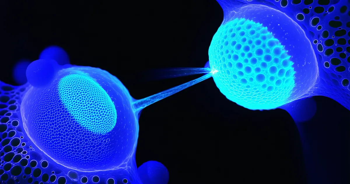
Transcytosis at the BBB is the process by which a substance is taken up from the luminal (blood-facing) side of the endothelial cell by endocytosis, transported across the cell in vesicles, and released on the abluminal (brain-facing) side by exocytosis. This is how the brain receives transferrin-bound iron, immunoglobulins, and certain peptide hormones from blood.
For RMT, the process begins with receptor binding at the luminal surface. The receptor is internalized with its ligand into a clathrin-coated vesicle or caveolar structure. The vesicle is transported to the abluminal membrane rather than routed to lysosomes (as would occur in a classical degradative endocytic pathway). At the abluminal surface, exocytosis releases the cargo into the brain interstitial space.
The distinction between productive transcytosis and degradative endocytosis is critical for LNP engineering. An LNP that binds a receptor and gets internalized but then traffics to lysosomes — where it would be degraded — does not achieve BBB crossing. The intracellular routing decision, not receptor binding per se, determines CNS delivery outcome. This is why simple uptake assays (which measure internalization but not transcytosis) overestimate BBB-crossing activity. An internalization efficiency of 70% measured by flow cytometry in an hCMEC/D3 culture does not mean 70% transcytosis — it means 70% of cells took up fluorescent signal, with no information about whether that signal was routed toward the abluminal side or toward degradative lysosomes.
Several intracellular factors govern the routing decision. Rab GTPase switching during endosomal maturation determines whether vesicles acquire the Rab7 or Rab11 identity that routes them to lysosomes versus recycling/transcytosis pathways. LNP surface properties can influence this switching — cationic particles tend to trigger Rab7 (degradative) routing, while particles engaging specific transcytosis receptors may bias toward Rab11 or transcytosis-specific Rab GTPases. These mechanisms are not fully characterized for LNP-sized particles, which is why empirical transwell transcytosis measurement remains the most reliable functional readout.
Transferrin Receptor 1 (TfR1, CD71)
TfR1 is the most extensively studied BBB transcytosis target for nanoparticle delivery. It is expressed at 5–10-fold higher density on brain endothelial cells compared to peripheral endothelium, providing a selectivity advantage for CNS targeting. Its endogenous ligand, transferrin, binds TfR1 with high affinity (Kd ~1 nM) and undergoes well-characterized clathrin-mediated endocytosis and transcytosis to supply the brain with iron necessary for neuronal metabolism and myelination.
Early TfR1 targeting work used anti-TfR1 antibodies (OX26, RI7217) conjugated to liposomes or nanoparticles and demonstrated statistically significant but modest CNS delivery improvements in rodents. Later work established that antibody affinity to TfR1 follows a counterintuitive optimization: very high-affinity TfR1 binders are less efficiently transcytosed than moderate-affinity binders, because high-affinity interactions favor retention in the endothelial cell rather than transcytosis to the abluminal side. The mechanistic interpretation is that high-affinity binding to TfR1 slows the rate of receptor dissociation in the endosomal compartment, and this sustained receptor engagement triggers lysosomal rather than transcytotic routing. This "affinity optimization" observation is mechanistically important for LNP surface engineering — it suggests that TfR1-targeting ligands should be designed for moderate affinity (in the 10–100 nM Kd range) rather than maximizing binding strength.
For LNPs, TfR1 targeting is achieved through surface display of TfR1-binding peptides or antibody fragments conjugated to the PEG-lipid corona. The density of targeting ligand matters: too low and receptor engagement is inefficient; too high and the particle may aggregate or trigger receptor crosslinking that routes to lysosomes rather than productive transcytosis. A key engineering consideration is that PEG-lipid with surface-conjugated targeting ligand and unconjugated PEG-lipid must be present at a ratio that provides steric shielding (from unconjugated PEG) while maintaining receptor accessibility of the targeting ligand. We typically work at 1–2 mol% functionalized PEG-lipid as a fraction of total PEG-lipid.
LRP1 (Low-Density Lipoprotein Receptor-Related Protein 1)
LRP1 is a large multi-ligand endocytic receptor expressed at high levels on brain endothelial cells. Its endogenous ligands include ApoE, ApoJ (clusterin), alpha-2-macroglobulin, lactoferrin, and receptor-associated protein (RAP). LRP1 is constitutively expressed on brain endothelium and mediates transcytosis of these ligands across the BBB — it is not significantly downregulated by iron status or common neuroinflammatory states in the same way that TfR1 expression can be modulated.
LNP targeting of LRP1 has been approached via ApoE-mimetic peptides displayed on the LNP surface. The mechanism mirrors the endogenous ApoE transcytosis pathway: the LNP, presenting an ApoE-mimetic ligand, binds LRP1 on the luminal surface, is internalized, and — in productive transcytosis — is transported to the abluminal side. In our hCMEC/D3 transwell data, LRP1 targeting alone achieves approximately 31% transcytosis efficiency at 4 hours, compared to approximately 8% for untargeted LNPs in the same model at matched particle concentrations. TEER of all monolayers was confirmed above 50 Ω·cm² prior to each experiment, and lucifer yellow paracellular leakage was below 1% to confirm monolayer integrity.
LRP1-targeted delivery faces a competitive binding issue that TfR1 targeting does not: serum ApoE circulates at approximately 30–60 µg/mL in adults, and alpha-2-macroglobulin circulates at even higher concentrations, creating significant competitive pressure for LRP1 binding sites. Surface ligand design must account for this — a peptide with affinity similar to serum ApoE will be substantially competed off by the endogenous ligand at physiological concentrations. This is why LRP1 targeting works better with designed peptides that have structural features distinguishing them from natural LRP1 ligands — particularly those that exploit the multi-domain architecture of LRP1, which has four ligand-binding clusters with partially distinct binding specificity — or with combinations that exploit avidity effects from multivalent LNP surfaces.
Dual Targeting: TfR1 + LRP1
The rationale for dual targeting is twofold. First, receptor expression levels vary across individual animals, disease states, and CNS regions — a single-receptor strategy is vulnerable to variable target expression. In neuroinflammatory conditions (relevant for HD, FA, and CLN3 Batten disease, where neuroinflammation accompanies neurodegeneration), TfR1 expression changes on activated microglia may complicate delivery targeting that relies solely on endothelial TfR1. Second, the two pathways appear to be partially independent; TfR1 and LRP1 internalize cargo through different endocytic routes (predominantly clathrin-mediated for TfR1 vs. caveolae/LRP-associated vesicles for LRP1), which may reduce competition for the same intracellular sorting machinery and allow additive transcytosis efficiency.
In our hCMEC/D3 transwell data, the dual-targeting formulation (TfR1 peptide + LRP1 ApoE-mimetic peptide, co-displayed on the LNP surface at optimized ligand density ratios) achieves 62% transcytosis efficiency at 4 hours — compared to 38% for TfR1 alone and 31% for LRP1 alone. The approximately 7.7-fold improvement over untargeted LNPs (8% efficiency) suggests that the two pathways contribute additively rather than redundantly in this in vitro model. We do not claim this reflects in vivo performance.
We are explicitly cautious about extrapolating transwell data directly to in vivo performance. The hCMEC/D3 model has a known limitation: TEER values in this cell line (typically 50–100 Ω·cm² in our cultures) are substantially lower than in vivo BBB TEER (1,500–8,000 Ω·cm²), meaning the model is more permeable than the actual BBB. This likely means our in vitro transcytosis numbers overestimate what we would observe in vivo. Primary human brain microvascular endothelial cell (hBMEC) co-culture models with tight junction-supporting pericytes and astrocyte end-feet achieve TEER values of 200–500 Ω·cm² and are more physiologically representative. We are developing data in those models. The hCMEC/D3 data is useful for formulation screening and comparison, not as a direct predictor of in vivo brain delivery.
What In Vitro Transcytosis Data Does and Does Not Mean
A transwell transcytosis assay measures the appearance of LNPs on the basolateral side of a cell monolayer over time. It is the most practical way to compare formulations at scale before committing to in vivo studies. But it abstracts away several features of the in vivo BBB that matter:
- Protein corona formation: In vivo, LNPs in circulation rapidly adsorb a protein corona from serum proteins within seconds of injection. This corona partially masks surface ligands and changes receptor binding characteristics. Transwell assays typically use diluted serum or serum-free conditions that do not accurately model corona formation. For CNS-targeted LNPs, the corona question is directly relevant — an LRP1-targeting ApoE-mimetic ligand that is buried under a serum albumin and fibrinogen corona will not reach LRP1 on the endothelial surface.
- Blood clearance and circulation time: Even a highly efficient BBB-crossing LNP must survive long enough in circulation to reach brain vasculature in sufficient concentration. PEG-lipid density, particle size, and zeta potential affect circulation half-life independently of transcytosis efficiency. An LNP with 60% in vitro transcytosis efficiency that is cleared from circulation in 5 minutes delivers less to the brain than one with 30% efficiency and a 4-hour half-life.
- CNS parenchymal diffusion after exocytosis: Transcytosis delivers LNPs to the brain interstitial space. From there, they must diffuse through extracellular matrix to reach neurons. Diffusion coefficients in brain ECM for LNP-sized particles are estimated in the range of 10⁻⁹ to 10⁻¹⁰ cm²/s — substantially lower than in free solution — meaning that even effective BBB crossing does not guarantee widespread neuronal distribution. LNPs that transcytose efficiently but diffuse poorly may achieve perivascular accumulation without meaningful neuronal transduction.
These caveats are not arguments against transwell models — they are essential for formulation comparison and candidate selection. They are arguments against treating transwell transcytosis data as equivalent to in vivo CNS delivery data. The two are related but not the same measurement.
Implications for LNP Surface Engineering
The mechanistic picture of BBB transcytosis places specific constraints on how LNPs should be engineered for CNS delivery.
Targeting ligand density must be titrated, not maximized. High ligand density may improve initial receptor binding but can impair intracellular routing toward productive transcytosis by triggering receptor crosslinking. We use a functionalized PEG-lipid conjugation approach that allows us to vary targeting ligand surface density independently of total PEG-lipid content, enabling systematic density titration experiments in hCMEC/D3 transwell assays before candidate formulations are taken to in vivo work.
Particle size should stay in the 70–100 nm range. Smaller particles below 50 nm may access more efficient transcytosis routes in some model systems, but are harder to produce at high loading efficiency for large cargo (base editor mRNAs, prime editor pegRNAs). Larger particles above 120 nm may engage multiple receptors simultaneously in ways that trigger lysosomal rather than transcytotic routing, or may be physically excluded from the clathrin-coated pit structures that initiate TfR1 endocytosis. Our current formulations target 83 nm z-average diameter with PDI below 0.15 as measured by DLS.
The LNP should not be cationic at physiological pH. Cationic particles trigger non-specific endocytosis and complement activation at the BBB endothelium, which routes cargo to degradative pathways rather than transcytosis and may also trigger endothelial inflammatory responses that compromise tight junction integrity — counterproductive for any delivery program. Ionizable lipid selection at physiological pH should maintain near-neutral or slightly anionic surface charge (our target: −5 to +5 mV zeta potential at pH 7.4 by electrophoretic light scattering).
The PEG-lipid density requires careful optimization for CNS applications specifically. Higher PEG density extends circulation time and reduces hepatic uptake — both desirable properties — but excessive PEG density sterically occludes targeting ligands from receptor engagement on the brain endothelial surface. CNS-targeted LNPs typically require lower PEG-lipid content (1.0–1.5 mol%) than the 2–2.5 mol% used in liver-optimized formulations to balance steric shielding with targeting ligand accessibility.
This is the mechanistic framework behind Biopathio's surface engineering approach. The full characterization data for our TfR1, LRP1, and dual-targeting formulations — including transwell transcytosis data, protein corona binding studies, and PEG density optimization results — is available on the BBB crossing platform page for research partners evaluating delivery vehicles for their CNS gene therapy programs.